Dental Practice · Oral Surgery
Extractions
Simple and surgical extractions, performed in-house using CBCT planning.
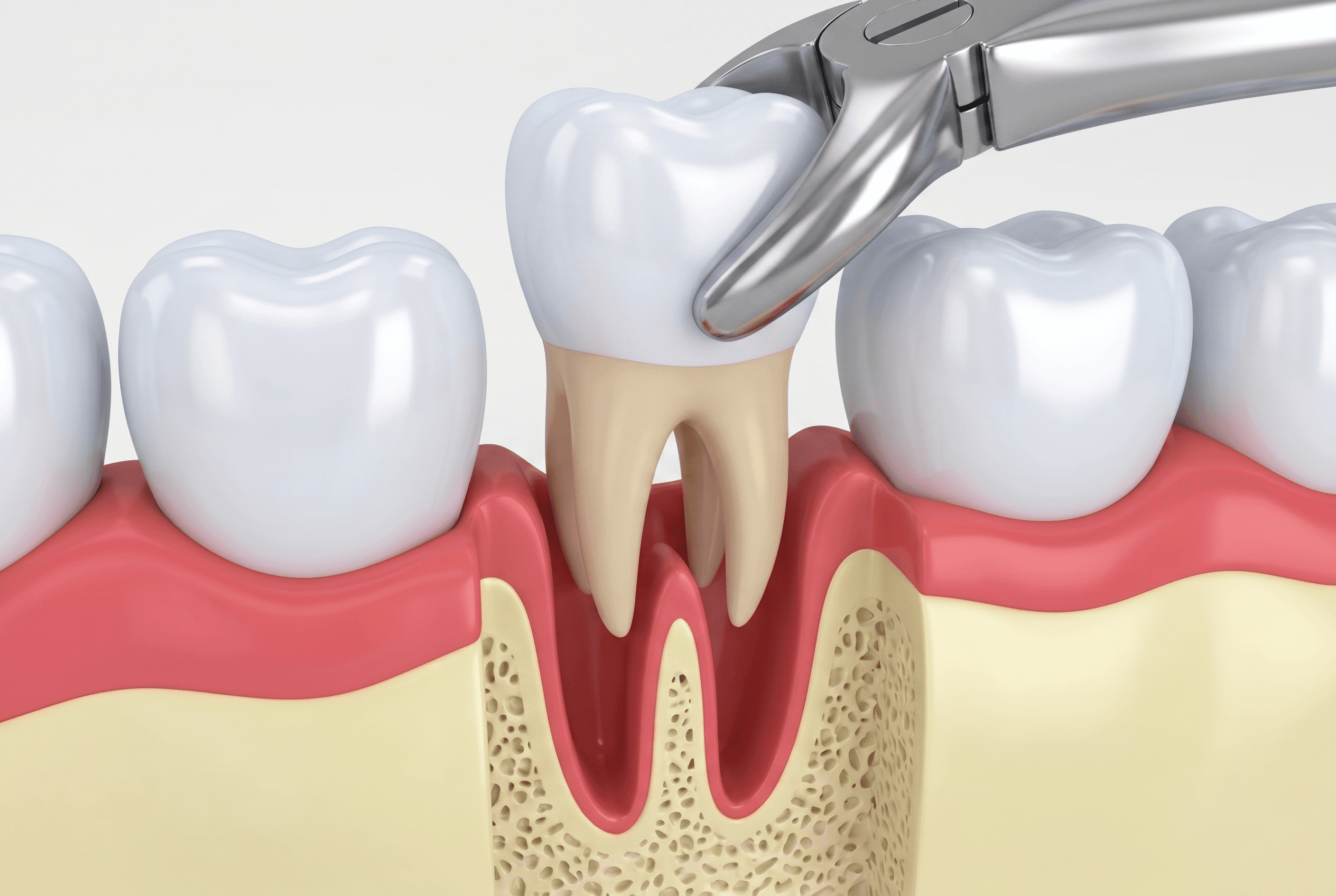
Our goal is to save every tooth we can. However, when extraction is necessary, we make that determination based on clinical evidence: extensive decay that leaves too little tooth structure for a restoration to hold, advanced periodontal disease with bone loss that cannot support the tooth's function, a fracture that extends below the bone level, symptomatic impaction, or orthodontic requirements. We explain the reasoning before we proceed. Most extractions can be done in our office under local anesthesia without a hospital referral or general sedation. Simple extractions (teeth with intact roots that can be loosened and removed in one piece) are straightforward. Surgical extractions involve sectioning the tooth, removing bone where necessary, or working around curved or fused root anatomy. We handle both. Before any surgical extraction, we capture a CBCT scan when the anatomy warrants it. The three-dimensional image from our iCAT FLX shows us the relationship between the roots and adjacent structures — the inferior alveolar nerve in the lower jaw, the maxillary sinus in the upper jaw, root curvature that a flat X-ray would underrepresent. That information changes the surgical approach and prevents avoidable complications. When bone grafting to preserve the site for future use is indicated, we discuss it before the extraction. Placing a graft at the time of extraction is significantly simpler and more effective than attempting augmentation later. Post-operative instructions are provided in writing at the appointment.